Lichen Sclerosus
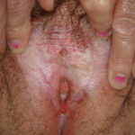
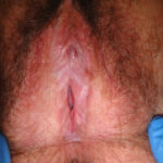
Below you will find a selection of clinical images portraying Lichen Sclerosus
1
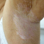
Axillary region showing a wide area of confluent porcelain-white papules and plaques with small ecchymoses (bruising) at 5 o'clock.
4
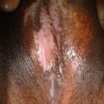
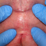
Pallor both sides anterior labia. Fusion anterior interlabial folds. Ulceration under clitoris.
7
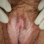
Pallor on vulva extending onto perineum. Loss of labia minora. Obscured clitoris, thickened white hood. Increased pigmentation around introitus.
10
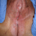
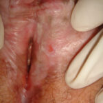
Loss of vulval structures. Very pigmented area near clitoris looks raised. Raised area left posterior labia. Scarred split perineal and perianal skin.
13
Loss of vulval structures with fusion.
Introitus fused.
2
5
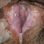
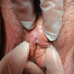
Pallor both labia minora. Obscured clitoris. Thickened clitoral hood. Loss of labial structures. Flattened interlabial sulci.
8
Thickened swollen clitoral hood. Obscured clitoris. Intense pallor on left labum minus with associated patch of pigmentation. Pallor and scarring extending to perineum and inner buttocks. Multiple ulcers including posterior fourchette.
11
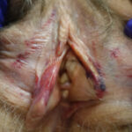
Labia minora conserved. Multiple ecchymoses on labia minora and labia majora. Swollen red right labum minus.
14
3
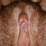
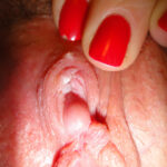
Fusion right lower clitoris. Thickened scarred pallor under hood.
6
Resorbed labia minora. Fusion across interlabial sulci. Obscured clitoris with fusion and thickening of hood. Pallor and areas of pigmentation. Scarring and one abrasion on left side. Reduced introitus.
9
Clitoris exposed by retraction of clitoral hood. Widespread pallor. Resorbed labia minora and flattened interlabial sulci. Increased pigmentation around introitus. Loss of elasticity.


