

1
Vulval Cancer and Lichen Sclerosus
A 2cm well demarcated nodular lesion (cancer) on the left labia with anatomical loss and clitoral hood fusion.
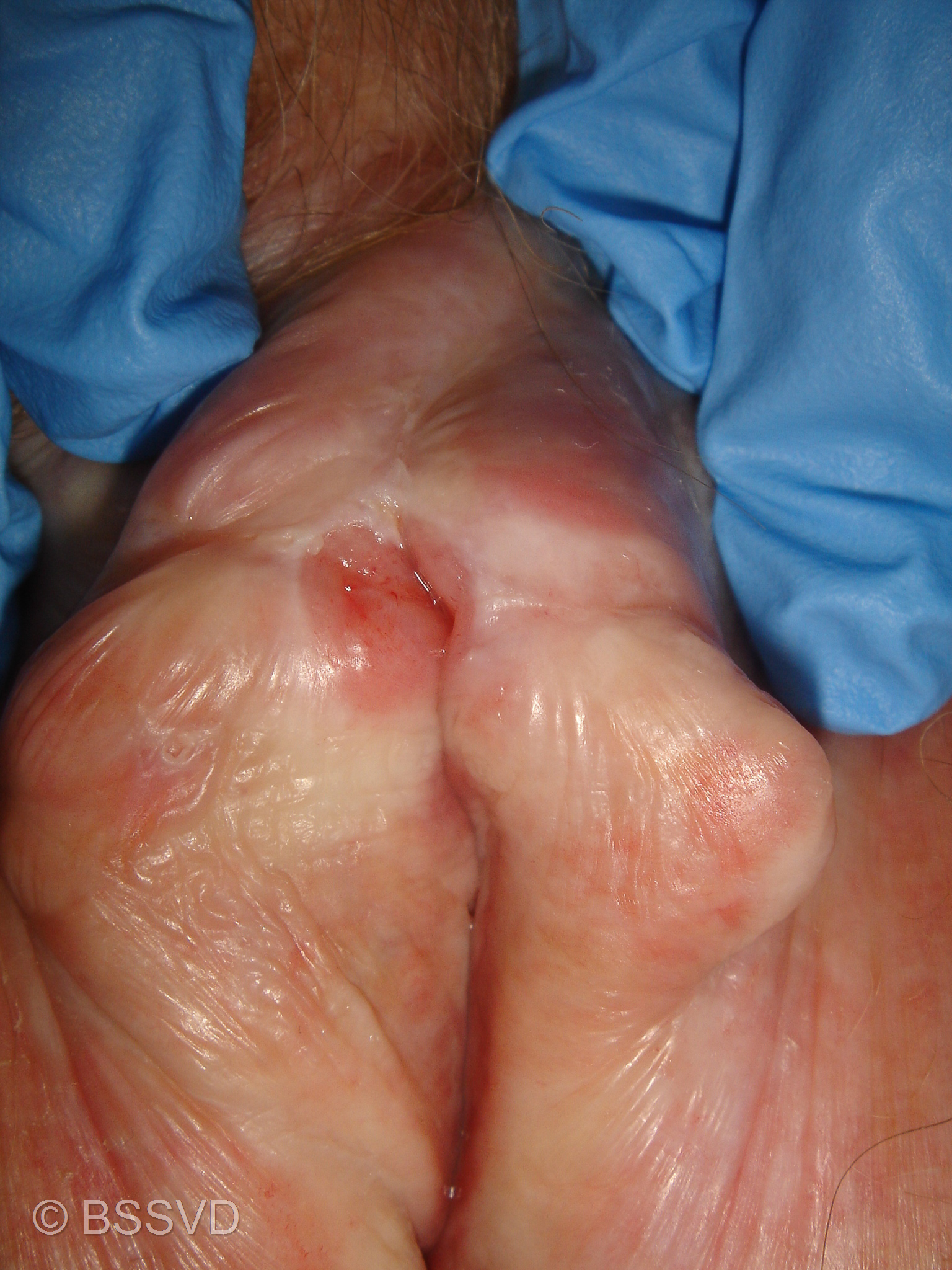
4
Perineal Vulval Cancer and Lichen Sclerosus and Planus
Widespread, whitening, ecchymosis and anatomical loss. Glazed erythema left side of the vulval vestibule. Perineal, well defined suspicious induration. Small posterior vaginal wall prolapse.

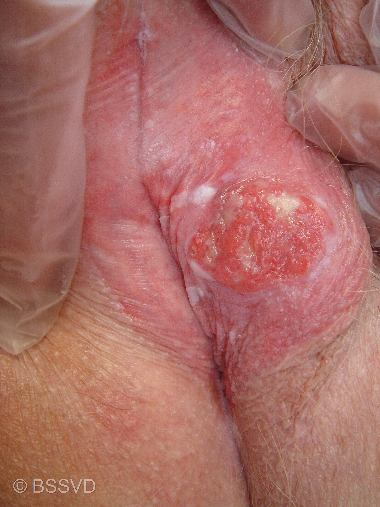
2
Vulval Cancer and Lichen Sclerosus
A 1cm well demarcated ulcerated lesion (cancer) under the clitoral hood with clitoral hood fusion and whitening.
5
Vulval Cancer and Lichen Sclerosus
A 2cm well demarcated nodular lesion (cancer) on the right labia with background lichen sclerosus.
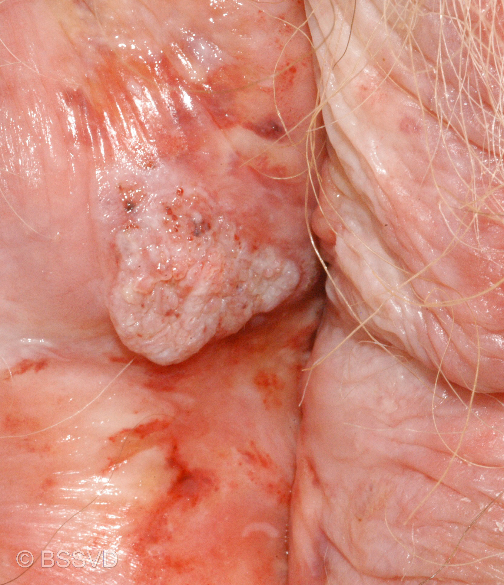
3
Vulval Cancer and Lichen Sclerosus
A small ulcerated lesion (cancer) on the lower aspect of the perineum close to the anus with background whitening.

6
Radiotherapy Effect on the Vulval post Vulvectomy for Vulval Cancer
Widespread scarring post vulvectomy, with telangiectasia over the vulva and mons pubis.
